Inside Robotic Knee Replacement — A Step by Step Look at How the Technology Works
The decision to undergo knee replacement surgery is a significant one, and uncertainty about what happens inside the operating theatre is one of the most common sources of anxiety for patients. When the procedure involves a robotic arm and computer screens, that unfamiliarity can feel even more intense.
This blog is designed to demystify the process. We will walk you through every phase of Fully Automated knee Replacement Surgery — from the first diagnostic scan to the moment you stand on your new knee — so that when the day comes, nothing feels unfamiliar.
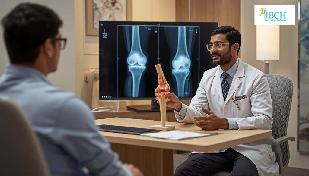
Step 1: Initial Consultation and Diagnostic Assessment
Everything begins with a thorough clinical consultation. Your Orthopaedic Surgeon will examine your knee, assess your range of motion, evaluate your gait, and review your medical history. Weight-bearing X-rays of the knee are taken in standing position to reveal the true extent of joint space narrowing, bone damage, and alignment deformity.
If robotic knee replacement is considered appropriate, a high-resolution CT scan of your knee is ordered. Unlike a standard X-ray, this CT scan captures a complete three-dimensional picture of your femur (thigh bone), tibia (shin bone), and patella (kneecap), including their exact dimensions, curvature, and current alignment.
Step 2: 3D Surgical Planning — The Digital Blueprint
The CT data is uploaded to the robotic system’s planning software. Here, a precise 3D model of your knee joint is generated — not a generic template, but a model that is unique to your anatomy. On this model, your surgeon plans the surgery in granular detail.
The planning process determines the exact size of the implant components, the angle and depth of each bone cut on the femur and tibia, the rotational alignment of the implant, and the projected post-operative limb alignment. The surgeon can virtually “test” different implant positions and predict how each will affect joint stability and range of motion.
This planning phase is one of the most powerful precision Robotic Knee Surgery benefits. It converts the operation from a procedure guided by intra-operative estimation into one guided by a validated, patient-specific blueprint.
Inside Robotic Knee Replacement — A Step by Step Look at How the Technology Works
Step 3: Day of Surgery — Anaesthesia and Registration
On the day of surgery, after anaesthesia is administered — typically regional anaesthesia (spinal or epidural) that numbs the lower body while keeping the patient comfortable — the surgical team begins the registration process.
Small optical tracking markers are placed on the bone near the knee joint. Infrared cameras in the operating theatre detect these markers, allowing the robotic system to track the exact real-time position of the femur and tibia. The system then overlays the pre-operative 3D plan onto the live anatomy, ensuring that the virtual blueprint and the physical knee are perfectly synchronised.
The surgeon verifies this registration by moving the knee through a series of positions and confirming that the on-screen model matches the actual joint movement. Only when registration accuracy is confirmed does the procedure advance.
Step 4: Robotic-Guided Bone Preparation
This is where the robotic system plays its most visible role. The surgeon uses a saw or burr held within the robotic arm to remove the damaged bone surfaces from the femur and tibia. The robotic arm constrains the cutting instrument to the exact pre-planned path — the surgeon guides the movement, but the robot ensures the boundaries are respected.
If the instrument approaches the edge of the planned cut zone, the robotic system provides haptic feedback (physical resistance) or automatically stops the blade. This prevents over-resection — removing more bone than necessary — which is one of the most common causes of implant malalignment in conventional surgery.
The cuts are precise, clean, and exactly as planned. This accuracy is the foundation of what the industry refers to when discussing minimally invasive robotic knee replacement: less bone is sacrificed, and the surrounding ligaments and soft tissues experience less disruption.
Step 5: Trial Fitting and Real-Time Balancing
After bone preparation, the surgeon places trial implant components on the prepared bone surfaces. The knee is taken through its full range of motion — bending, straightening, rotating — while the robotic system displays real-time data on joint gap measurements, ligament tension, and overall alignment.
This step is critical. A well-balanced knee — one where the inner and outer ligaments bear load evenly through the full range of motion — is the difference between a knee that feels natural and one that feels “off.” In conventional surgery, this balancing relies entirely on the surgeon’s tactile assessment. With robotic assistance, the surgeon has objective, quantified data supplementing their clinical feel.
If the data reveals any imbalance, the surgeon can make micro-adjustments — a fractional change in bone resection depth or implant rotation — before committing to the final implant. This iterative, data-driven approach is why robotic knee replacements consistently achieve better functional scores in published clinical studies — and why long-lasting knee implants with robotics are not just a claim but a measurable clinical outcome.
Step 6: Final Implant Placement and Closure
Once the surgeon is fully satisfied with the trial fit and balance, the final implant components are placed. Depending on the implant system and the patient’s bone quality, the components may be cemented (fixed with bone cement) or cementless (press-fitted for biological integration). The surgeon performs a final assessment of stability and range of motion, then closes the wound in layers.
Step 7: Post-Operative Recovery and Early Mobilisation
Recovery begins almost immediately. Within a few hours of surgery, the physiotherapy team assists the patient in sitting up, performing ankle pumps and gentle knee bends, and — for many patients — standing with support. Supervised walking with a walker typically starts the next morning.
The emphasis on early mobilisation is deliberate. Movement promotes blood circulation (reducing the risk of blood clots), prevents stiffness, and kick-starts the muscle re-education process. Because the robotic approach preserves more muscle and ligament tissue, patients generally tolerate early movement with noticeably less discomfort than they expect.
Most patients are discharged within two to four days with a structured home physiotherapy protocol. Follow-up visits are scheduled at regular intervals — typically at two weeks, six weeks, three months, six months, and one year — to monitor healing, implant stability, and functional progress.
What Makes This a Top Robotic Joint Replacement Center in West Bengal
The combination of advanced robotic technology, a dedicated high-volume surgical team, and a structured end-to-end patient programme is what distinguishes a leading centre from one that merely owns a robotic system. Salt Lake, Kolkata, is home to orthopaedic centres that have built their robotic knee replacement programmes around this integrated approach — from the very first consultation through long-term post-operative follow-up.
With robotic joint replacement surgery in West Bengal becoming increasingly accessible, patients no longer need to delay a decision that could transform their quality of life. Understanding the process removes uncertainty and builds the confidence needed to take the next step toward better mobility.
If you would like to understand how robotic knee replacement can specifically help with your condition, a one-on-one consultation with an experienced orthopaedic specialist is the best place to start. JBCH Hospital in Salt Lake, Kolkata, is a centre patients across West Bengal can explore for robotic knee replacement.